
Understanding Abdominal Wall Hernia Symptoms and What to Expect with Ultrasound Diagnosis
- kerryrob
- Feb 5
- 3 min read
Abdominal wall hernias are a common health issue that many people experience but often misunderstand. Recognizing the symptoms early and knowing how ultrasound helps in diagnosis can make a significant difference in managing this condition effectively. This post explains what an abdominal wall hernia is, the symptoms you might notice, and how ultrasound imaging plays a crucial role in confirming the diagnosis and guiding treatment.

What Is an Abdominal Wall Hernia?
An abdominal wall hernia happens when an internal part of the body, usually a section of the intestine or fatty tissue, pushes through a weak spot or tear in the muscles of the abdominal wall. This creates a visible bulge or lump under the skin. Hernias can develop in different areas of the abdomen, such as the groin (inguinal hernia), around the belly button (umbilical hernia), or at the site of a previous surgical incision (incisional hernia).
The weakness in the abdominal wall can be present from birth or develop over time due to factors like heavy lifting, persistent coughing, obesity, or previous surgeries. Understanding the symptoms and seeking timely diagnosis helps prevent complications such as pain, obstruction, or strangulation of the herniated tissue.
Common Symptoms of Abdominal Wall Hernia
Symptoms vary depending on the hernia size, location, and whether it is reducible (can be pushed back into the abdomen) or incarcerated (trapped outside the abdominal wall). Here are the most common signs to watch for:
Visible bulge or lump: Often the first noticeable symptom, especially when standing or straining.
Pain or discomfort: This may increase with physical activity, coughing, or lifting heavy objects.
A feeling of pressure or weakness: Some people describe a dragging sensation in the affected area.
Swelling or tenderness: The skin over the hernia may become red or tender if complications arise.
Nausea or vomiting: These symptoms can indicate a more serious problem like bowel obstruction.
If you experience sudden severe pain, redness, or vomiting, seek medical attention immediately as these may signal strangulation, which requires urgent treatment.
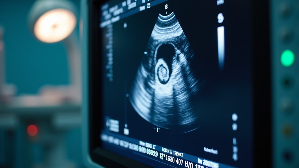
How Ultrasound Helps Diagnose Abdominal Wall Hernias
Ultrasound is a safe, non-invasive imaging technique that uses sound waves to create real-time images of the abdominal wall and its contents. It is often the first choice for diagnosing hernias because it provides clear visualization without radiation exposure.
What to Expect During an Ultrasound Exam
The procedure usually takes 15 to 30 minutes.
You will lie on an examination table while a technician applies a gel to your abdomen.
A handheld device called a transducer moves over the skin to capture images.
You may be asked to cough or strain to make the hernia more visible.
The exam is painless and does not require special preparation.
Benefits of Ultrasound in Hernia Diagnosis
Detects hernias not visible on physical exam: Small or deep hernias can be hard to feel but show up clearly on ultrasound.
Differentiates hernias from other lumps: Ultrasound helps distinguish hernias from cysts, tumors, or swollen lymph nodes.
Assesses hernia contents and size: Knowing what tissue is involved guides treatment decisions.
Monitors hernia progression: Follow-up ultrasounds track changes over time or after surgery.

What Happens After Diagnosis
Once a hernia is confirmed by ultrasound, your healthcare provider will discuss treatment options based on the hernia’s size, symptoms, and your overall health. Options include:
Watchful waiting: Small, painless hernias may only require monitoring.
Lifestyle changes: Avoiding heavy lifting, managing weight, and treating chronic cough can reduce strain on the abdominal wall.
Surgical repair: Recommended for larger or symptomatic hernias to prevent complications. Surgery can be open or laparoscopic.
Ultrasound may be used again after surgery to check for recurrence or complications.
When to See a Doctor
If you notice a new lump or bulge in your abdomen or groin, especially if it causes discomfort or pain, schedule an evaluation. Early diagnosis with ultrasound can prevent serious issues and help you understand what you are experiencing.


Comments