
Steps and Importance of Appendix Imaging Procedure
- Chib Onwunaka

- Apr 7
- 4 min read
When it comes to diagnosing abdominal pain, especially in the lower right side, the appendix often becomes the focus. Understanding the appendix imaging procedure can make a significant difference in timely and accurate diagnosis. I want to walk you through the essential steps involved in this process and explain why it’s so important for your health or the care of those you assist.
What Is the Appendix Imaging Procedure?
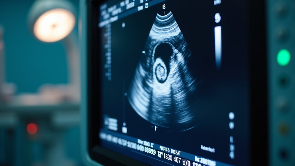
The appendix imaging procedure is a non-invasive diagnostic test used to visualize the appendix and surrounding tissues. It helps detect inflammation, infection, or other abnormalities that might indicate appendicitis or other conditions. This procedure is typically done using ultrasound technology, which uses sound waves to create images of the inside of the body.
Ultrasound is preferred because it is safe, painless, and does not expose you to radiation. It’s especially useful for children, pregnant women, and individuals who need a quick and reliable diagnosis.
Step-by-Step Guide to the Appendix Imaging Procedure
If you or someone you care for needs an appendix imaging procedure, knowing what to expect can ease any anxiety. Here’s a clear breakdown of the steps involved:
Preparation
You may be asked to avoid eating or drinking for a few hours before the test. This helps reduce gas in the intestines, which can interfere with the images.
Positioning
You will lie down on an examination table, usually on your back. The technician might ask you to change positions slightly to get better views of the appendix.
Applying Gel
A water-based gel is applied to the lower right side of your abdomen. This gel helps the ultrasound probe make better contact with your skin and improves image quality.
Scanning
The technician moves the ultrasound probe gently over the area. You might feel slight pressure but no pain. The probe sends sound waves into your body and captures the echoes to create images.
Image Review
The technician captures several images from different angles. These images are then reviewed by a radiologist or your healthcare provider to look for signs of inflammation or other issues.
Results and Follow-Up
Results are usually available quickly. If the appendix appears inflamed or abnormal, your healthcare provider will discuss the next steps, which may include further testing or treatment.
This straightforward process is designed to be as comfortable and efficient as possible, ensuring you get the answers you need without unnecessary stress.
Why the Appendix Imaging Procedure Matters
The importance of this procedure cannot be overstated. Appendicitis is a common emergency that requires prompt diagnosis and treatment. Delays can lead to complications such as a ruptured appendix, which can be life-threatening.
Here’s why the appendix imaging procedure is crucial:
Early Detection
It helps identify appendicitis before it worsens, allowing for timely medical intervention.
Non-Invasive and Safe
Unlike CT scans, ultrasound does not expose you to radiation, making it safer for repeated use and for vulnerable populations.
Accurate Diagnosis
It provides clear images that help differentiate appendicitis from other causes of abdominal pain, such as ovarian cysts or gastrointestinal issues.
Cost-Effective
Ultrasound is generally more affordable and accessible, which aligns with the goal of providing vital diagnostic imaging to everyone, regardless of insurance status.
Supports Treatment Decisions
The images guide healthcare providers in deciding whether surgery or conservative management is appropriate.
Understanding these benefits can give you peace of mind and confidence in the diagnostic process.

Tips for a Smooth Appendix Imaging Procedure
To get the most accurate results and a comfortable experience, consider these practical tips:
Follow Pre-Test Instructions
If fasting or other preparations are required, stick to them closely.
Wear Comfortable Clothing
Loose-fitting clothes make it easier to access the abdomen and reduce discomfort.
Communicate with the Technician
Let them know if you feel any pain or discomfort during the scan.
Stay Still During the Scan
Movement can blur the images, so try to relax and remain as still as possible.
Ask Questions
Don’t hesitate to ask about the procedure or what the images show. Understanding the process helps reduce anxiety.
By preparing well and staying informed, you can help ensure the procedure goes smoothly and yields the best possible results.
How Accessible Appendix Imaging Procedure Can Be
Access to quality diagnostic imaging should never be a barrier to good health. That’s why facilities like VirtuScan Imaging are committed to making sonography services affordable and accessible across the DFW Metroplex. Whether you are at a medical clinic, a skilled nursing center, or receiving care at home, you can expect a personal touch and professional service.
If you or someone you care for needs an appendix ultrasound, you can trust that the procedure will be handled with care, respect, and expertise. This approach ensures that everyone, regardless of insurance status, can get the vital diagnostic imaging they need.
Taking the time to understand the steps and importance of the appendix imaging procedure empowers you to make informed decisions about your health or the care you provide. Remember, early and accurate diagnosis is key to effective treatment and peace of mind.


Comments